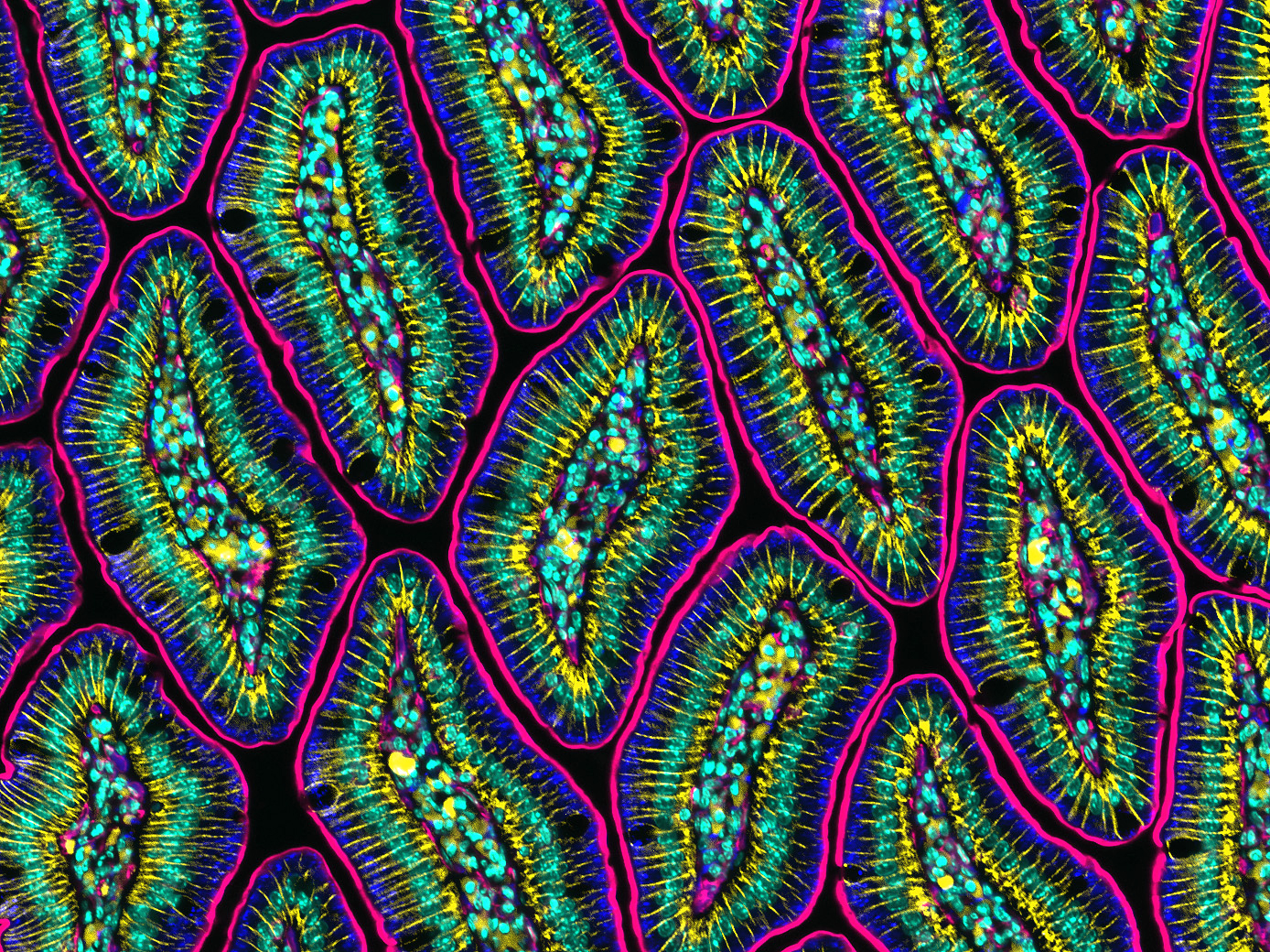
Mouse intestine

Murine small intestine showing various cellular villi features. Antibodies were used to identify the apical membrane (pink), the lateral membrane between cells (yellow), and the nuclei (teal). Fluorescence microscopy. Image by Amy Engevik, Medical University of South Carolina, Charleston, SC