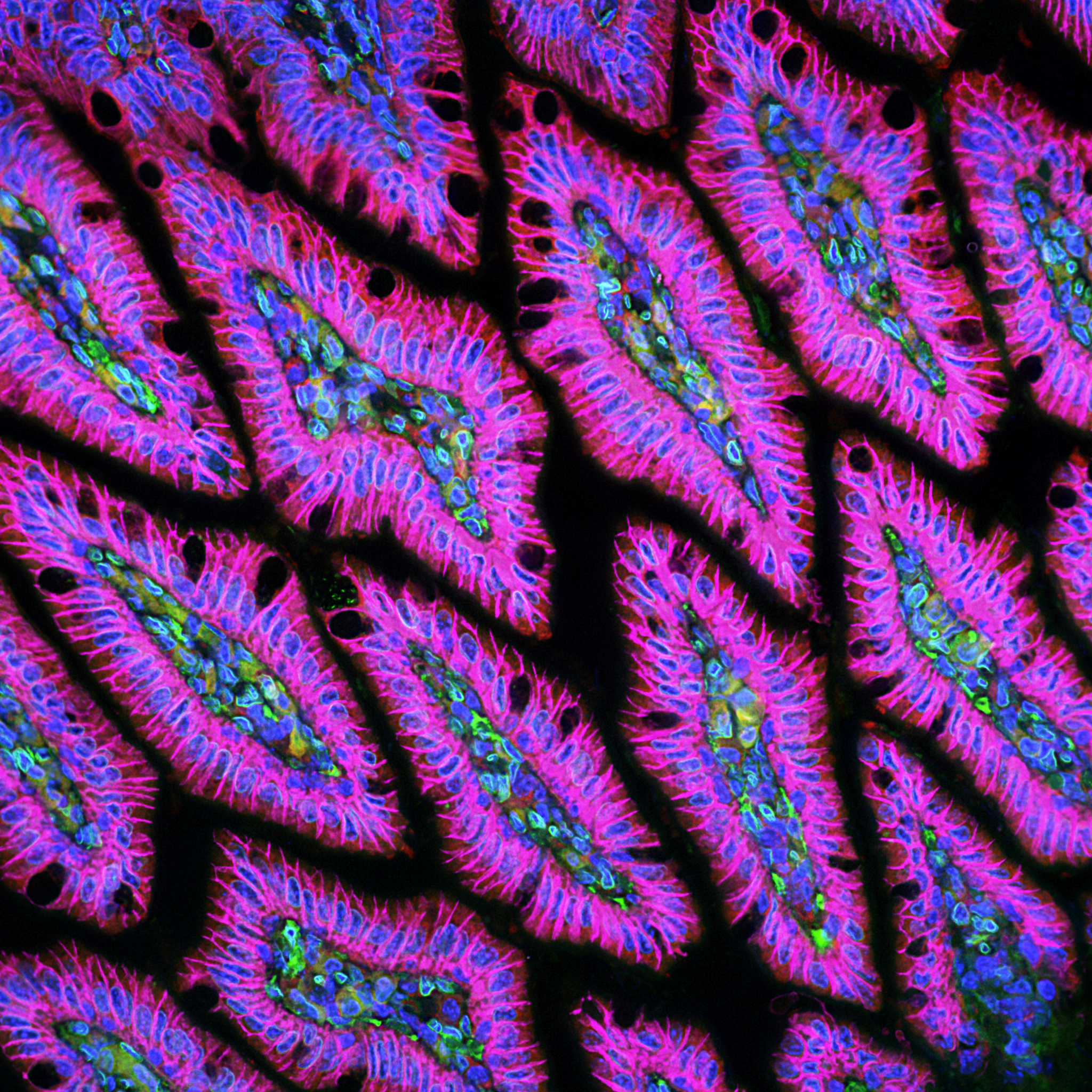
Mouse colon

Imunofluorescence image of mouse colon tissue section showing the complex spatial structure of nuclei, cytoplasm, and cell membranes within the tissue. Shades of the colors blue, green, red, and magenta indicate the nuclei, nuclei membranes, cytoplasm, and cell membranes, respectively. Fluorescence microscopy. Image by Bogdan Kochetov, University of Pittsburgh, Pittsburgh, PA