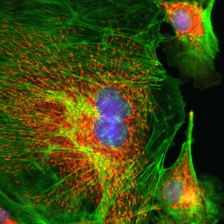
Epifluorescence imaging of rat endothelial cells
Rat endothelial cells were fixed on a glass slide and marked with fluorescent dyes. Nuclei were stained with DAPI (blue), actin filaments with phalloidin (green), and mitochondria with MitoTracker™. Sample from Anja Kraemer of Boehringer Ingelheim Pharma GmbH. Images acquired with a confocal Raman microscope.

Submitted by: Damon Strom, WITec GmbH, Ulm, Germany